Posts

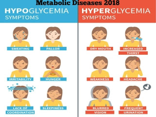
At what time diabetes patients can drink milk?

Image
A change in breakfast schedule may give benefits for the management of type 2 diabetes . A study inspected the impacts of devouring high-protein milk at breakfast on blood glucose levels and satiety after breakfast and after a moment dinner. Milk devoured with breakfast cereal diminished postprandial blood glucose concentration compared with water, and high dairy protein concentration decreased postprandial blood glucose concentration compared with ordinary dairy protein concentration. The high-protein treatment moreover diminished appetite after the lunch compared with the low-protein proportionate. Metabolic diseases are on the rise universally, with type 2 diabetes and weight as driving concerns in human well-being. In this way, there's impulse to create dietary procedures for the risk reduction and management of obesity and diabetes to engage buyers to progress their individual well-being. In this randomized, controlled, double-blinded study, the analysts in

Diabetes and Frozen Shoulder

Image
Among the numerous complications frequently connected with diabetes, one of the foremost mystifying and painful is the frozen shoulder. Adhesive capsulitis , the condition’s biological term, is a pathological condition of the shoulder joint which causes the painful and gradual loss of motion. The connective tissue of the shoulder joint inflames and stiffens, causing chronic pain, limiting mobility and disturbing sleep. The associated pain varies between sharp stings that radiate through the bicep to dull ache. Link between diabetes and frozen shoulder: There’s no conclusive connect link between the shoulder condition and the disease. Few studies indicate frozen shoulder is caused by glycosylation of the collagen in the shoulder joint.  What Happens to the Shoulder? Adhesive capsulitis is characterized by three stages, in spite of the fact that the severity and length of the stages may vary from individuals to individuals. ·          Stage One: Freezing - The

Bioartificial Pancreas – A recent advancement in the treatment of Diabetes

Image
A bioartificial pancreas —a device that supports and encapsulates islets of Langerhans— replaces the beta cells and islets which were destroyed by type 1 diabetes. Implanted in the peritoneal cavity or under the skin, it contains approximately a million islets. It responds to changing blood glucose levels by releasing hormones, mainly insulin. Every bioartificial pancreas is manufactured from non-living and living components. The living component is the islets, which secrete insulin concurring to typical physiology by sensing glucose levels. The non-living component shields the islets from the diabetic’s body and its destructive immune mechanisms, however grants the islets inside to thrive. There are several types of bioartificial pancreas which includes microencapsulated , or coated islets are the first-generation bioartificial pancreas. Their advantage is that nutrients can effectively move into the islets and insulin can move out. Among the downsides is that they are diff

Diabetes and Cholesterol Metabolism

Image
The maintenance of blood glucose in a limit run requires tight coupling among circulating insulin secretion, proinsulin biosynthesis and nutrients. Glucose, the most physiological stimulus for insulin secretion, controls insulin release through activities on so-called triggering and amplifying pathways in the β-cell. The ATP-sensitive K+ (K+-ATP) channels are key players within the activating pathway. Their activity is directed by the mitochondrial oxidative phosphorylation of glucose metabolites, which comes about in an increased cytosolic ATP to ADP ratio, hence restraining potassium efflux through the channel, depolarizing the plasma membrane, and opening voltage-dependent L-type calcium channels, which increments cytosolic calcium and leads to exocytosis of insulin. In addition to the triggering pathway, the β-cell displays pathways downstream to the K + -ATP channel, which increases insulin secretion to abdicate physiological levels of the hormone in response to glucose. T

Learning about Familial Hypercholesterolemia

Image
Familial hypercholesterolemia is a genetic disorder. It is caused by a defect on chromosome 19. It causes LDL (Low Density Lipoprotein) cholesterol level to be very high. The deformity makes the body incapable to remove LDL (bad) cholesterol from the blood. This results in a high level of LDL in the blood.  The condition is typically passed down through families in an  autosomal dominant  way. This means you merely need to get the abnormal gene from one parent in order to acquire the disease.  In uncommon cases, a child may acquire the gene from both parents. When this happens, the increase in cholesterol level is much more severe. The risk for heart attacks and heart disease are high, even in childhood. High cholesterol became a very common medical condition, but it's frequently the result of unhealthy lifestyle choices, and thus treatable and preventable. With familial hypercholesterolemia, a person's chance of high cholesterol is higher because a defect (mutation

Hypoglycaemia in Diabetes

Image
In diabetes, hypoglycaemia for the most part emerges as a result of pharmacologic treatment that causes blood glucose levels to drop underneath the normal range. Whereas mild hypoglycaemia usually resolves with incite ingestion of carbohydrates, more often reduced glucose levels can impede cognitive function, and even threaten life. Nocturnal hypoglycaemia poses a specific challenge because the sleeping patient isn't in a position to intervene and numerous episodes are asymptomatic. Repeated exposure to nocturnal hypoglycaemia can limit counterregulatory mechanisms, with possibly serious clinical consequences. Hypoglycaemia in diabetes typically happens as a result of therapeutic treatment, particularly with insulin, glinides or sulfonylureas (SUs). Risk factors for hypoglycaemia includes Aggressive treatment of glycaemia, HbA1C < 6.5%* (except in youth), High glucose variability (in older adults), Long duration of diabetes, Renal impairment, Duration of insulin therap

Stem Cell – Is a treatment for diabetes?

Image
Stem cells are the cells that have a potential for creating an entire human body. A   stem cell  has the ability to divide, and the divided new  cell  has the potential either to remain a  stem cell  or become another type of  cell  with a more specialized function, such as a brain cell , or a red blood  cell . Researchers have found ways to isolate them outside the human body, concentrate in a laboratory and embed back. Stem cells treatment require administration of isolated cells in the damaged area, wherein they can divide in the harmed area, adapt the properties of inhabitant stem cells and start some of the misplaced functions that have been compromised by the disease or injury. Various researches are accessible proposing in vitro differentiation of stem cells into insulin-producing beta cells. These cells can as well offer assistance in creating a microenvironment due to initiate secretion of different immune cells to counteract autoimmunity of the individual.  Developing